Technology

List Of Food Items Rich In Vitamin C

List Of Food Items Rich In Vitamin C

orange slices

Marcus Clackson / Istock

You may be surprised to find which food ranks number one. Hint: It's not oranges.

orange slices

Marcus Clackson / Istock

1 of 11

The Health Benefits of Vitamin C

Vitamin C helps the body maintain healthy tissues and a strong immune system, and it aids in the absorption of iron. Vitamin C is also considered a powerful ally if you're trying to kick a cold.

The recommended daily allowance for adult women is 75 milligrams and for adult men it's 90 milligrams. Get your vitamin C naturally with these top 10 food sources, according to the USDA's Dietary Guidelines for Americans.

guava

Hugo Chang/ Istock

2 of 11

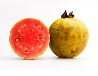
Guava

One half cup of the fruit contains 188 milligrams of vitamin C and only 56 calories.

red sweet pepper

Matjaz Boncina / Istock

3 of 11

Red Pepper

You might instantly think of citrus when you think of immune-boosting vitamin C, but sweet red peppers offer a solid dose of the nutrient, too. One half cup of the pepper contains 142 milligrams of vitamin C and 20 calories. If you cook the veggie, a half cup offers 116 milligrams.

kiwi

Potapova Valeriya / Istock

4 of 11

Kiwi

One medium kiwi contains 70 milligrams of the nutrient and 46 calories.

orange

IStock

5 of 11

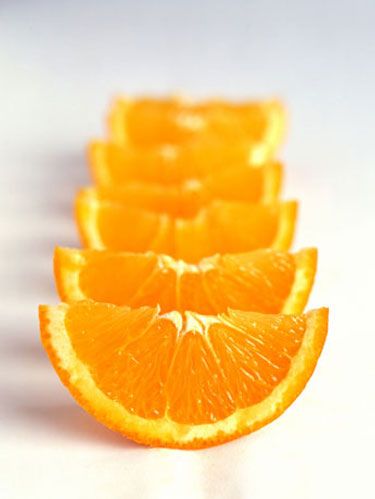
Orange

One medium orange contains 70 milligrams of vitamin C and 62 calories. If you prefer it in liquid form, 3/4 cup of orange juice contains 61 to 93 milligrams of vitamin C and 79 to 84 calories.

green pepper

Lucas Cornwell / Istock

6 of 11

Green Pepper

Green peppers offer 60 milligrams of vitamin C per half cup, with only 15 calories. When cooked, the vegetable provides 51 milligrams of vitamin C.

grapefruit juice

Lucas Cornwell / Istock

7 of 11

Grapefruit Juice

One half of a grapefruit has 39 milligrams of vitamin C and 52 calories.

For more of the vital nutrient, go for the liquid: Three fourths of a cup of grapefruit juice provides 50 to 70 milligrams of vitamin C and 71 to 86 calories.

vegetable juice cocktail

Image Source / Getty Images

8 of 11

Vegetable Juice

Drinking your vegetables provides plenty of nutrients, including a vitamin C punch (and, if your veggie juice is tomato-based, a healthy dose of lycopene, a disease-fighting phytonutrient). You'll get 50 milligrams of vitamin C and 34 calories in 3/4 cup of juice.

strawberries

Enrico Fianchini/ Istock

9 of 11

Strawberries

Strawberries are full of fiber and antioxidants, including vitamin C. A half cup of strawberries contains 49 milligrams of the vitamin and 27 calories.

brussels sprouts

Christine Balderas / Istock

10 of 11

Brussels Sprouts

This veggie may not be the most kid-friendly choice, but it is always a healthy pick — packed with both 48 milligrams of vitamin C, 300 micrograms of vitamin K, and just 28 calories.

cantaloupe

Elena Schweitzer/ Istock

11 of 11

Cantaloupe

One quarter of a medium cantaloupe has 47 milligrams of vitamin C and 51 calories.

Advertisement - Continue Reading Below

This content is created and maintained by a third party, and imported onto this page to help users provide their email addresses. You may be able to find more information about this and similar content at piano.io

List Of Food Items Rich In Vitamin C

Source: https://www.goodhousekeeping.com/health/diet-nutrition/g1926/top-sources-vitamin-c-44102808/

Keto Sources Of Vitamin C

Keto Sources Of Vitamin C

Foods high in vitamin C have long been hailed as immunity-boosting powerhouses and essential for inclusion in your day-to-day diet. According to the Office of Dietary Supplements, vitamin C is vital to protect against illness and disease as, in the body, it acts as an antioxidant helping to protect cells from the damage caused by free radicals.

What is vitamin C?

Also known as ascorbic acid or nicknamed 'the immunity vitamin', the water-soluble nutrient is most commonly found in fruit and veg.

What does vitamin C do?

Vitamin C has a number of purposes in the body, explains nutritional therapist Paula Werrett. 'It acts as an antioxidant, helping to protect cells from free radicals created through normal energy production and ingested through foods and from sunlight and pollution in the environment.'

And that's not all. 'The body also requires vitamin C to produce collagen, a protein needed for the maintenance of health for bones, cartilage, gums, teeth and skin to help wounds heal. Finally, vitamin C is also extremely important to help with the absorption of iron-rich foods and helps the immune system to manage the disease', she shares. Not bad.

Ensuring you absorb enough iron from your diet is especially important if you exercise regularly. Working on the reg although good for you can actually contribute to iron deficiency, so giving your body a helping hand if you're a fan of a good sweat sesh definitely won't do any harm.

What else is vitamin C good for?

Vitamin C is also needed to prevent deficiency diseases such as scurvy, and is helpful for the maintenance of good energy and metabolism and for healthy skin, cartilage, bones and teeth, she expands.

But it is also relevant to coronavirus . 'Vitamin C has been shown to help shorten the length and severity of respiratory infections, such as colds.' This means, in short, that eating enough regularly can can help you bounce back from common cold and flu symptoms more quickly, she explains.

So what's the link between vitamin C and Coronavirus?

Doctor Aragona Giuseppe, GP and medical advisor at Prescription Doctor, says it's important to clarify the vit C's role in dealing with COVID-19: 'There is no medical or scientific evidence to suggest that a certain food or supplement can help prevent a person catching COVID-19'.

vitamin c foods vegetables

yulkapopkova

Dr Giuseppe explains that because vitamin C is well-known as an immunity boosting vitamin many people assume that it can help fight off coronavirus. 'While vitamin C could help to reduce or get rid of any symptoms, there is no evidence to suggest that vitamin C alone will fight off the virus. Every body is different and is being affected by the virus differently', he continues.

There is no evidence to suggest that vitamin C alone will fight off the virus

However, Heather Rosa (@IONHRosa), the Dean of the Institute for Optimum Nutrition, disagrees, sharing that a study suggests early and high intravenous doses of Vitamin C could help to fight the coronavirus disease. 'A recent Medicine in Drug Discovery study led by Virginia Commonwealth University found that patients who received intravenous vitamin C spent significantly fewer days in intensive care and hospital overall. On average, the vitamin C group spent three fewer days in the ICU and a week less in the hospital overall.'

vitamin c foods closeup of a cheerful young couple picking some fruit and veggies from the fridge to make some healthy breakfast on sunday morning shot from inside the working fridge

gilaxia

Plus, she shares that a group of hospitals in New York are treating their most critical COVID-19 patients with high doses of vitamin C.

Only time will tell the true impact vitamin C can have on the virus as scientists, doctors and experts continue to work towards a vaccine and potential counters. Whatever the effect of the vitamin, Giuseppe advises listening to government guidelines, staying at home and making a few small lifestyle tweaks, if you don't already do these things, to ensure you keep yourself (and your immune system) in tip top shape.

  • Eat plenty of nutrient dense foods (Giuseppe advises fruit, fish, healthy oils, protein and vegetables)
  • Stay hydrated with eight glasses of water a day
  • Get moving and use your hour a day outside wisely
  • Be mindful of your alcohol intake
  • Maximise your sleep and aim for eight hours a night (if you're struggling with this, read our guide to clean sleeping).

    However, experts do maintain that eating a diet high in antioxidants and whole, non-processed foods generally will support bodily functioning and, in turn, potentially assist your immune system in fighting off viruses.

    How much vitamin C do I need per day?

    'The NHS recommends we get 40 mg a day', says Rosa. 'Anything more than 1,000 mg a day and you may experience stomach pain and diarrhoea', she warns.

    The World Health Organisation guidelines state similar, recommending 45mg of vitamin C per day or 300mg per week from your diet.

    To put that into context, a small orange contains around 51g of vitamin C. You know what they say—an orange a day…

    However, Werrett believes that the sky is the limit with vitamin C consumption—within reason. 'The RDA of 40mg is the minimum amount you need to consume to prevent deficiency disease, such as scurvy. Much higher levels, however, may be optimal, with the Department of Health and Social Care suggesting up to 1,000mg daily for adults is unlikely to cause harm. Recent research trials show that still larger doses of 3,000 to 5,000mg a day (or even higher) may be needed for optimal effects in protection against viruses.'

    What are the best vitamin C foods?

    As mentioned, vitamin C is found predominantly in plants, with oranges the most well known natural source.

    • Acerola cherries
    • Blackcurrants
    • Broccoli
    • Brussel Sprouts
    • Chilli peppers
    • Guavas
    • Kale
    • Kiwis
    • Kakadu plum
    • Lemons
    • Lychees
    • Mustard
    • Oranges
    • Papayas
    • Parsley
    • Red peppers
    • Spinach
    • Strawberries
    • Sweet yellow peppers
    • Thyme

      As you can see while oranges are high in vit C, they're not the only fruit or veg.

      What is the best source of vitamin C?

      Fun fact: 100g of red pepper has 4.5 times the amount of vitamin C than an orange and a certain type of plum has over ten times. Scroll to see how 100g of orange stacks up against 100g of its rivals, in order from the highest:

      • Kakadu plum - 7000 mg per 100g veg
      • Red Peppers - 242.5 mg per 100g veg
      • Kale - 120 mg per 100g veg
      • Kiwi - 92.7 mg per 100g veg
      • Broccoli - 89.2 mg per 100g veg
      • Brussels Sprouts - 85 mg per 100g veg
      • Strawberries - 58.8 mg per 100g veg
      • Orange - 53.2 mg1 per 100g veg.

        Can you overdose on Vitamin C?

        Generally speaking, no, although if you consume too much, the majority will be excreted in your urine, shares nutritionist Lily Soutter (@lily_soutter_nutrition). 'You may also experience symptoms such as stomach pain, diarrhoea and flatulence if you ingest 1g per day or more.'

        Plus, Rosa stressed that high doses of ascorbic acid (vitamin C) can wear away tooth enamel. She recommends washing your mouth out with a little water after consumption.

        If you have type 1 diabetes, G6DP deficiency or hyperoxaluria, Rosa also advises that consuming too much vitamin C could be damaging or detrimental for reasons independent to each condition.

        Keen to read up on vitamin C skincare and how it could perk up your skin? You're in the right place—read the WH guide to learn more about that.

        Should I take a vitamin C supplement?

        'Eating a healthy diet including plenty of Vitamin C rich foods is important for overall health. Extra vitamin C from supplements, however, may be needed to support the immune system further at times of stress or to further optimise the immune system,' shares Werrett. Essentially, you should consider your exercise levels, exposure to pollution and general diet.

        This content is imported from {embed-name}. You may be able to find the same content in another format, or you may be able to find more information, at their web site.

        If you are worried about deficiencies, turning to vitamin C supplements can be an easy and effective way of ensuring you're meeting your RDA.

        Vitamin C Supplements

        Boots VITAMIN C 1000 mg Effervescent tablets

        Boots VITAMIN C 1000 mg Effervescent tablets

        boots pharmaceuticals boots.com

        £4.00

        Solgar Natural Cranberry with Vitamin C Vegi Capsules

        Solgar Natural Cranberry with Vitamin C Vegi Capsules

        Solgar hollandandbarrett.com

        £14.99

        Holland & Barrett Iron & Vitamin C 100 Tablets

        Holland & Barrett Iron & Vitamin C 100 Tablets

        hollandandbarrett.com

        £3.29

        Boots Vitamin C 30 Gummies

        Boots Vitamin C 30 Gummies

        boots pharmaceuticals boots.com

        £4.00


        Disclaimer: The information in this story is accurate as of the publication date. While we are attempting to keep our content as up-to-date as possible, the situation surrounding the coronavirus pandemic continues to develop rapidly, so it's possible that some information and recommendations may have changed since publishing. For any concerns and latest advice, visit the World Health Organisation . If you're in the UK, the National Health Service can also provide useful information and support, while US users can contact the Center for Disease Control and Prevention .

        Cut through the noise and g et practical, expert advice, home workouts, easy nutrition and more direct to your inbox. Sign up to t he WOMEN'S HEALTH NEWSLETTER .

        This content is created and maintained by a third party, and imported onto this page to help users provide their email addresses. You may be able to find more information about this and similar content at piano.io

        Keto Sources Of Vitamin C

        Source: https://www.womenshealthmag.com/uk/food/healthy-eating/a32223259/vitamin-c-foods/

        Is 1000mg Of Vitamin C Too Much For A Teenager

        Is 1000mg Of Vitamin C Too Much For A Teenager

        Photo Courtesy: KARRASTOCK/Moment/Getty Images

        Nearly 2 million American adults each year are diagnosed with cancer. If you receive a diagnosis, your schedule can quickly start to fill up with doctor's appointments, medical procedures and pharmacy visits. Creating a checklist of things to do can help keep you busy and feeling more in control. But acknowledging the effects a cancer diagnosis can have on your emotions — and healthily managing those emotions — is almost as important.

        Doctors and psychologists now recognize that healing improves after a diagnosis when both our physical and emotional needs are met, says Niki Barr, Ph.D., a psychotherapist at the Center for Cancer and Blood Disorders in Texas, who consults with medical doctors, extended families and caregivers about emotionally coping with a cancer diagnosis.

        Learning you have cancer is a stressful experience, and it's important to acknowledge and cope with that stress. Fully understanding how your emotional needs can change after you receive a cancer diagnosis can go a long way in helping you and your family heal. The tips here can help you support your mental health as you navigate your diagnosis and treatment.

        Photo Courtesy: burakkarademir/E+/Getty Images

        Upon hearing your diagnosis, you might experience grief, fear or even a feeling of denial until some time has passed. "These are all normal and emotionally healthy responses, but it's all too easy to spiral out of control with fear in the beginning," says Barr. Fortunately, these tips can help you manage the anxiety and fear that come along with learning about a cancer diagnosis.

        Lessen the Impact of Anxious Thoughts

        Start by writing your thoughts down on note cards or in a journal. Identify the first one that's leading you to feel uneasy. For example, you might write down something like "I'm afraid of my hair falling out." Then, move onto the subsequent fear and write it down. That might be something like "People will treat me differently if they see I have cancer."

        When you've identified most of your anxious thoughts, go back to the first one and write something new on the card that can help ease your stress. It should be a thought that's confident and empowering. For example, suppose you're worried about your hair falling out. In that case, a positive view could be: "I've been looking forward to getting a new hairstyle anyway." When you're feeling nervous, read the more positive strategies, says Barr.

        Work On Your Internal Dialogue

        It can also help to defuse all those "What if?" questions you might ask yourself, such as "What if my cancer has spread?" or "What if the treatment doesn't work?" One scary question tends to lead to another and often turns into full-fledged anxiety. Try to focus on those things you have the power to improve on your own. The next time you start asking yourself the what-ifs, substitute the upsetting ideas with this one: "Is this thought helping me or hurting me?" You can also ask, "Is this thought moving me forward or backward?" Your answers might surprise you, and they can help you move away from the thoughts that are holding you back.

        Get Grounded

        Interrupting periods of anxiety by focusing on small details around you can help you shift your perspective away from negativity and towards the present moment. "Look at the beautiful color of the walls in the room you're in; look at the person you're talking to, the clothes [they're] wearing," Barr suggests. Becoming very present and focused on physical details nearby helps soothe sudden anxious thoughts. Turn your focus towards absorbing the colors, smells, people and each new sound around you. Build those sensations up very clearly in your mind. You can use this technique as a distraction tool the next time you're waiting for a medical procedure or want a diversion from your thoughts.

        Meditate to Music

        Research shows that 15 to 30 minutes of both guided imagery and soothing tunes can alleviate deep feelings of stress about a cancer diagnosis. The Cancer Treatment Centers of America (CTCA) reviewed 30 clinical cancer trials that analyzed more than 1,890 cancer patients. They found that music therapy can have a beneficial effect on anxiety, pain, mood, quality of life, heart rate, respiratory rate and blood pressure.

        The CTCA also offers many classes and therapies to help people relax, reduce stress and improve their quality of life as part of an extensive mind-body medicine program. Many centers provide calming background music during healing therapies.

        What Are Your Next Steps?

        Photo Courtesy: Peathegee Inc/Getty Images

        You have a cancer diagnosis: Now what? Learning how to clear your mind and focus on positive thoughts is a helpful step in the right direction. Here are other soul-soothing strategies you can try.

        Start Journaling and Reflecting

        Research published in the Journal of Clinical Oncology has shown that expressing your innermost feelings can reduce stress and have a range of other emotional and social benefits. Researchers aren't sure why putting thoughts down on paper is effective. Still, it allows you to process complex emotions and help you chart a way forward, whether you've been diagnosed with cancer or are taking care of someone who has.

        Exercise When Possible

        Exercising for 2.5 hours per week can help you beat symptoms of depression and fatigue. Among the nation's millions of cancer survivors, there are hints — but not proof yet — that active exercisers may lower their risk of their cancer coming back.

        The American College of Sports Medicine hosted a medical panel of cancer specialists to evaluate this exercise evidence. It issued guidelines suggesting that cancer patients and survivors should exercise for about 30 minutes most days of the week. This exercise should be enjoyable to you but also build up a sweat. When patients and their family caregivers exercised together, research found, everyone was more likely to stick with the fitness regimen, boost their physical stamina and experience less emotional strain.

        Strengthen Your Social Support System

        Connecting with others who've been through this kind of emotional diagnosis already can be a source of comfort and support. Learn more about online communities and your local chapters and support groups that meet up. The American Cancer Society also suggests attending one-on-one professional therapy or its "I Can Cope" online support groups to learn more.

        Resource Links:

        Find Local Cancer Support Programs | Cancer Support Groups

        Strategies Used in Coping With a Cancer Diagnosis Predict Meaning in Life for Survivors

        Coping Well with Advanced Cancer: A Serial Qualitative Interview Study with Patients and Family Carers

        Coping with cancer

        Coping Attitudes of Cancer Patients and Their Caregivers and Quality of Life of Caregivers

        MORE FROM SYMPTOMFIND.COM

        Is 1000mg Of Vitamin C Too Much For A Teenager

        Source: https://www.symptomfind.com/health/tips-coping-with-cancer-diagnosis?utm_content=params%3Ao%3D740013%26ad%3DdirN%26qo%3DserpIndex

        How Do I Give My Guinea Pig Vitamin C

        How Do I Give My Guinea Pig Vitamin C

        Style

        These Are The 6 Best Vitamin C Serums For Brighter, Clearer Skin

        Amazon/Bustle

        There's never a bad time to amp up your skin care regimen's sun protection defenses. Whether you're looking to keep your skin safe from future signs of sun damage or help reduce the appearance of damage that's already been done (aka sun spots), incorporating one of the best vitamin C serums into your beauty routine can help.

        In an ingredients list, you'll most likely find vitamin C listed as l-ascorbic acid, which experts agree is the most effective form of vitamin C, along with tetrahexyldecyl ascorbate. You'll also want to pay attention to the concentration level of the vitamin C, as something between 10 and 20 percent is said to be the most stable, and therefore, the most effective when it comes to protecting the skin and improving its overall tone and texture.

        While you can certainly apply your vitamin C serum at night, to reap its full sun-protecting benefits, apply it in the morning after cleansing, and before you moisturize and layer on your SPF (which, yes, you still need to wear). You'll also want to be careful about using your vitamin C serum after any cleansers, treatments, or moisturizers that contain salicylic acid, glycolic acid, or retinol, as this can heighten common side effects that come with these ingredients, like burning and redness (especially if you have sensitive skin).

        It's also worth noting that vitamin C is a delicate ingredient because it degrades from sun exposure. Because of this, you'll want to store your serum away from the sunlight and be on the lookout for any changes in its color, texture, or smell. A serum packaged in a darker bottle is also ideal, as it helps minimize the product's exposure to sunlight.

        Ahead, you'll find six of the best vitamin C serums that are all Amazon user-approved to brighten, protect, and firm your skin.

        1

        The Best Serum For Vitamin C Newbies

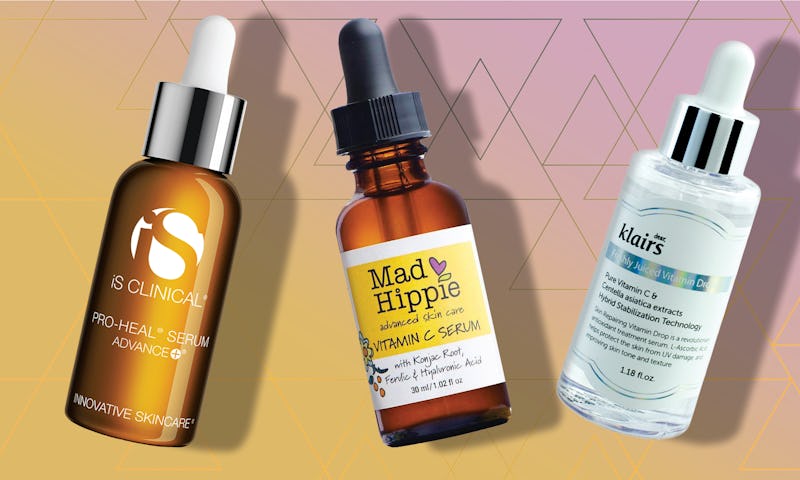
        This K-beauty favorite serum contains a mild dose of 5 percent vitamin C to allow your skin to accustom itself with the active ingredient. While that's still enough to notice the brightening and skin tone-evening benefits, the smaller percentage means its also gentler and less likely to irritate skin. Reviewers love its lightweight texture and say a little goes a long way, so you only need a couple of drops. Some have even had luck with using it on their bodies to help fade unwanted scars.

        Klairs Freshly Juiced Vitamin C Serum is also an Amazon-user favorite with hundreds of five-star reviews. "I used this product for a few months and then switched to one with a higher percentage of Vitamin C only to find that it didn't work nearly as well," writes one reviewer. "I'm back to using Klairs and am so impressed by the positive change I see in my skin when I use it — it's brighter, smoother, and more even — even though it's a mild product."

        The only downside with this serum is that, since it comes in a clear bottle, you'll want to be extra careful with storing it somewhere away from the sun.

        2

        The Best Vitamin C Serum For Sensitive Skin

        If you have sensitive skin, you might have luck with the low-concentration Klairs option, above. But it does contain a few ingredients that some particularly sensitive skin types negatively react to, like propylene glycol. For a super safe, all natural pick, try this all-natural pick from Mad Hippie. It uses vitamin C in the form of sodium ascorbyl phosphate, which is said to cause less irritation and reduce the risk of oxidation. Mad Hippie formulated this serum with other complementary ingredients, like vitamin E and ferulic acid, which both improve and protect the skin from damaging free radicals. And ferulic acid also helps stabilize both vitamins C and E, so that they're even more effective. To keep skin moisturized, hyaluronic acid is included in the formula, while konjac root and chamomile extract work to protect and soothe the skin. There's also a dose of clary sage, which acts as a natural toner and astringent.

        "I have very sensitive skin and normally I cannot use vitamin C serums at all," writes a reviewer. "This one does not sting or burn at all." Another user notes, "Within a week or so, I saw a smoother texture to my skin. By the end of the bottle, my skin looked smoother, brighter, and three darker spots on my face [from sun damage] were at least 80 percent lighter."

        3

        A Vitamin C Serum That Produces An Instant Glow

        Yes, all vitamin C serums brighten the skin with continued use, but The Body Shop Vitamin C Skin Boost Instant Smoother also leaves skin glowing directly after you apply it, thanks to the tiny mineral glowing particles it contains. Long-term brightening, protecting, and collagen-boosting effects come from ascorbic acid and the vitamin C-packed camu camu berries that The Body Shop has hand harvested by local collectors on the riverbanks of the Amazon River. Vitamin E provides the skin with more protection against environmental aggressors as it softens, and Community Fair Trade Brazil nut oil further moisturizes the skin. Additionally, dimethicone and glycerin give the serum a smooth texture that helps to blur pores and even out skin tone, making this a great primer, too.

        4

        The Best Vitamin C Serum For Acne-Prone Skin

        This serum might be a bit of a splurge, but its powerful formula means serious results. The brand works with a team of assorted experts to develop each product, including geneticists, biologists, and physicians. For their iS Clinical Pro-Heal Serum Advance+, they developed a vitamin C combo that works for all skin types, including acne-prone. Time-released vitamin C means prolonged antioxidant protection, while vitamins E and A contribute even more antioxidant benefits. iS Clinical included inflammation-reducing zinc sulfate, blemish-preventing kojic acid, and nourishing olive leaf extract to combat signs of acne while helping to prevent future breakouts.

        5

        The Best Drugstore Vitamin C Serum

        For a more affordable option, e.l.f's Beauty Shield Vitamin C Pollution Prevention Serum costs less than $20. Despite its low price, the serum still contains your vitamin C, as well as vitamin E and pomegranate extract, which provides even more vitamin C benefits. You'll also notice glycerin and hyaluronic acid on the ingredients list to leave the skin smooth and moisturized. It's even cruelty-free and vegan, and formulated without parabens, sulfates, or phthalates.

        6

        The Best Vitamin C Booster Powder

        If you're into DIY beauty (or you just want to customize your vitamin C application), a vitamin C booster powder, like the Resurrection Beauty L-Ascorbic Acid Powder, is the perfect choice. This jar solely contains vitamin C in the form of l-ascorbic acid, which is meant to be dissolved in water or witch hazel before being added into your favorite hydrating serum. It can even be added to your nightly moisturizer, sunscreen, or body lotion. Because you're mixing this yourself, you can start with a lower concentration of vitamin C, like 5 percent, and work your way up to 20 percent (the maximum amount recommended) as your skin learns to tolerate it.

        Many Amazon reviewers share the different ways they use this powder to make custom treatments. "How terrific to find cosmetic grade vitamin C to make my own solution," one raves. "All skin care experts claim this is the best for anti-aging, however very unstable. This fixes the problem and at an reasonable price. I just mix small batches to last for a week. And in just a few weeks I can see the difference in my skin." Another recommends mixing it with aloe-vera for a skin-soothing and brightening mask.

        Bustle may receive a portion of sales from products purchased from this article, which was created independently from Bustle's editorial and sales departments.

        How Do I Give My Guinea Pig Vitamin C

        Source: https://www.bustle.com/p/the-6-best-vitamin-c-serums-16980515

        Glossier Vitamin C Reddit

        Glossier Vitamin C Reddit

        Photo Courtesy: KARRASTOCK/Moment/Getty Images

        Nearly 2 million American adults each year are diagnosed with cancer. If you receive a diagnosis, your schedule can quickly start to fill up with doctor's appointments, medical procedures and pharmacy visits. Creating a checklist of things to do can help keep you busy and feeling more in control. But acknowledging the effects a cancer diagnosis can have on your emotions — and healthily managing those emotions — is almost as important.

        Doctors and psychologists now recognize that healing improves after a diagnosis when both our physical and emotional needs are met, says Niki Barr, Ph.D., a psychotherapist at the Center for Cancer and Blood Disorders in Texas, who consults with medical doctors, extended families and caregivers about emotionally coping with a cancer diagnosis.

        Learning you have cancer is a stressful experience, and it's important to acknowledge and cope with that stress. Fully understanding how your emotional needs can change after you receive a cancer diagnosis can go a long way in helping you and your family heal. The tips here can help you support your mental health as you navigate your diagnosis and treatment.

        Photo Courtesy: burakkarademir/E+/Getty Images

        Upon hearing your diagnosis, you might experience grief, fear or even a feeling of denial until some time has passed. "These are all normal and emotionally healthy responses, but it's all too easy to spiral out of control with fear in the beginning," says Barr. Fortunately, these tips can help you manage the anxiety and fear that come along with learning about a cancer diagnosis.

        Lessen the Impact of Anxious Thoughts

        Start by writing your thoughts down on note cards or in a journal. Identify the first one that's leading you to feel uneasy. For example, you might write down something like "I'm afraid of my hair falling out." Then, move onto the subsequent fear and write it down. That might be something like "People will treat me differently if they see I have cancer."

        When you've identified most of your anxious thoughts, go back to the first one and write something new on the card that can help ease your stress. It should be a thought that's confident and empowering. For example, suppose you're worried about your hair falling out. In that case, a positive view could be: "I've been looking forward to getting a new hairstyle anyway." When you're feeling nervous, read the more positive strategies, says Barr.

        Work On Your Internal Dialogue

        It can also help to defuse all those "What if?" questions you might ask yourself, such as "What if my cancer has spread?" or "What if the treatment doesn't work?" One scary question tends to lead to another and often turns into full-fledged anxiety. Try to focus on those things you have the power to improve on your own. The next time you start asking yourself the what-ifs, substitute the upsetting ideas with this one: "Is this thought helping me or hurting me?" You can also ask, "Is this thought moving me forward or backward?" Your answers might surprise you, and they can help you move away from the thoughts that are holding you back.

        Get Grounded

        Interrupting periods of anxiety by focusing on small details around you can help you shift your perspective away from negativity and towards the present moment. "Look at the beautiful color of the walls in the room you're in; look at the person you're talking to, the clothes [they're] wearing," Barr suggests. Becoming very present and focused on physical details nearby helps soothe sudden anxious thoughts. Turn your focus towards absorbing the colors, smells, people and each new sound around you. Build those sensations up very clearly in your mind. You can use this technique as a distraction tool the next time you're waiting for a medical procedure or want a diversion from your thoughts.

        Meditate to Music

        Research shows that 15 to 30 minutes of both guided imagery and soothing tunes can alleviate deep feelings of stress about a cancer diagnosis. The Cancer Treatment Centers of America (CTCA) reviewed 30 clinical cancer trials that analyzed more than 1,890 cancer patients. They found that music therapy can have a beneficial effect on anxiety, pain, mood, quality of life, heart rate, respiratory rate and blood pressure.

        The CTCA also offers many classes and therapies to help people relax, reduce stress and improve their quality of life as part of an extensive mind-body medicine program. Many centers provide calming background music during healing therapies.

        What Are Your Next Steps?

        Photo Courtesy: Peathegee Inc/Getty Images

        You have a cancer diagnosis: Now what? Learning how to clear your mind and focus on positive thoughts is a helpful step in the right direction. Here are other soul-soothing strategies you can try.

        Start Journaling and Reflecting

        Research published in the Journal of Clinical Oncology has shown that expressing your innermost feelings can reduce stress and have a range of other emotional and social benefits. Researchers aren't sure why putting thoughts down on paper is effective. Still, it allows you to process complex emotions and help you chart a way forward, whether you've been diagnosed with cancer or are taking care of someone who has.

        Exercise When Possible

        Exercising for 2.5 hours per week can help you beat symptoms of depression and fatigue. Among the nation's millions of cancer survivors, there are hints — but not proof yet — that active exercisers may lower their risk of their cancer coming back.

        The American College of Sports Medicine hosted a medical panel of cancer specialists to evaluate this exercise evidence. It issued guidelines suggesting that cancer patients and survivors should exercise for about 30 minutes most days of the week. This exercise should be enjoyable to you but also build up a sweat. When patients and their family caregivers exercised together, research found, everyone was more likely to stick with the fitness regimen, boost their physical stamina and experience less emotional strain.

        Strengthen Your Social Support System

        Connecting with others who've been through this kind of emotional diagnosis already can be a source of comfort and support. Learn more about online communities and your local chapters and support groups that meet up. The American Cancer Society also suggests attending one-on-one professional therapy or its "I Can Cope" online support groups to learn more.

        Resource Links:

        Find Local Cancer Support Programs | Cancer Support Groups

        Strategies Used in Coping With a Cancer Diagnosis Predict Meaning in Life for Survivors

        Coping Well with Advanced Cancer: A Serial Qualitative Interview Study with Patients and Family Carers

        Coping with cancer

        Coping Attitudes of Cancer Patients and Their Caregivers and Quality of Life of Caregivers

        MORE FROM SYMPTOMFIND.COM

        Glossier Vitamin C Reddit

        Source: https://www.symptomfind.com/health/tips-coping-with-cancer-diagnosis?utm_content=params%3Ao%3D740013%26ad%3DdirN%26qo%3DserpIndex

        Eskimo Vitamin C

        Eskimo Vitamin C

        Dear Cecil:

        Why didn't Eskimos get scurvy before citrus was introduced to their diet? They have a traditional diet of almost entirely meat and fish. Where did they get their vitamin C?

        Kevin Carson, via the Internet

        Illustration by Slug Signorino

        Cecil replies:

        This calls to mind a question I've dealt with before: Why do the Eskimos (or Inuit, as those in Canada and Greenland generally prefer to be called) stay there? It turns out that the people of the north have a highly evolved physiology that makes them well suited to life in the arctic: a compact build that conserves warmth, a faster metabolism, optimally distributed body fat, and special modifications to the circulatory system. One marvels at the adaptability of the human organism, of course, but still one has to ask: Wouldn't it have been easier just to move to San Diego?

        Much of what we know about the Eskimo diet comes from the legendary arctic anthropologist and adventurer Vilhjalmur Stefansson, who made several daredevil journeys through the region in the early 20th century. Stefansson noticed the same thing you did, that the traditional Eskimo diet consisted largely of meat and fish, with fruits, vegetables, and other carbohydrates — the usual source of vitamin C — accounting for as little as 2 percent of total calorie intake. Yet they didn't get scurvy.

        Stefansson argued that the native peoples of the arctic got their vitamin C from meat that was raw or minimally cooked — cooking, it seems, destroys the vitamin. (In fact, for a long time "Eskimo" was thought to be a derisive Native American term meaning "eater of raw flesh," although this is now discounted.) Stefansson claimed the high incidence of scurvy among European explorers could be explained by their refusal to eat like the natives. He proved this to his own satisfaction by subsisting in good health for lengthy periods — one memorable odyssey lasted for five years — strictly on whatever meat and fish he and his companions could catch.

        A few holdouts didn't buy it. To settle the matter once and for all, Stefansson and a colleague lived on a meat-only diet for one year under medical supervision at New York's Bellevue Hospital, starting in February 1928. The two ate between 100 and 140 grams of protein a day, the balance of their calories coming from fat, yet they remained scurvy free. Later in life Stefansson became a strong advocate of a high-meat diet even if you didn't live in the arctic; he professed to enjoy improved health, reduced weight, etc, from meals consisting of coffee, the occasional grapefruit, and a nice steak, presumably rare. Doesn't sound half bad, and one might note that until recently the Inuit rarely suffered from atherosclerosis and other Western ailments.

        Vitamin C can be found in a variety of traditional Eskimo/Inuit staples, including the skin of beluga whales (known as muktuk), which is said to contain as much vitamin C as oranges. Other reported sources include the organ meats of sea mammals as well as the stomach contents of caribou. You're thinking: It'll be a mighty cold day in the arctic before they catch me eating the stomach contents of caribou. Indeed, you have to wonder whether the Inuit really ate such stuff either, since Stefansson describes it being fed to dogs.

        Other aspects of the arctic diet also remain controversial. For example, some say the Eskimos could get vitamin C from blueberries during the summer months, while others say you'd be lucky to find enough berries to cover a bowl of Rice Chex. I say let's not sweat the details of the menu, which varied from region to region anyway. We know Eskimos got enough vitamin C in their traditional diet to survive because obviously they did. Now it's academic — most arctic natives live in villages and get their vitamin C from OJ and Juicy Juice, just like you and I.

        Oh, and for all you vegetarians who've seen the error of your ways and were thinking of adopting the Inuit diet — think twice about the raw meat thing. Vitamin C might not a problem, but E. coli might.

        Questions we're still thinking about

        Dear Cecil:

        I read in a recent magazine that the ringworm parasite cannot stand anesthesia. So if and when a victim of ringworm is put under for any medical purpose, the ringworm has been known to use any means of escape possible, and it would not be unusual to see quite large infestations evacuating through a patient's nostrils almost immediately upon anesthetizing. True? And are there any documented incidences of this? Can you find pictures?

        — Morbid Curiosity in Providence

        Cecil replies:

        Definitely an interesting visual, Morb. Unfortunately (for you), ringworm is a fungus, not a critter. Guess you're stuck with tapeworms, huh?

        Cecil Adams

        Send questions to Cecil via cecil@straightdope.com.

        Eskimo Vitamin C

        Source: https://www.straightdope.com/21343509/traditionally-eskimos-ate-only-meat-and-fish-why-didn-t-they-get-scurvy

        Does Vitamin C Produce Collagen

        Does Vitamin C Produce Collagen

        Abstract

        Vitamin C (Vit C) benefits to human skin physiology notably by stimulating the biosynthesis of collagen. The main cutaneous collagens are types I and III, which are less synthesized with aging. Vit C is one of the main promotors of collagen formation but it poorly bypasses the epidermis stratum corneum barrier. To address this challenge, we developed a lipophilic version of Vit C for improving skin diffusion and delivery. Vit C was covalently conjugated to squalene (SQ), a natural lipid of the skin, forming a novel Vit C–SQ derivative suitable for cream formulation. Its biological activity was investigated on human whole skin explants in an ex vivo model, through histology and protein and gene expression analyses. Results were compared to Vit C coupled to the reference lipophilic compound palmitic acid, (Vit C–Palmitate). It was observed that Vit C–SQ significantly increased epidermal thickness and preferentially favored collagen III production in human skin after application for 10 days. It also promoted glycosaminoglycans production in a higher extent comparatively to Vit C–Palmitate and free Vit C. Microdissection of the explants to separate dermis and epidermis allowed to measure higher transcriptional effects either in epidermis or in dermis. Among the formulations studied, the strongest effects were observed with Vit C–SQ.

        Introduction

        Vitamin C (Vit C) or L-ascorbic acid has important physiologic effects on skin, including promotion of collagen biosynthesis, inhibition of melanogenesis, prevention of radiation-induced damages and acceleration of wound healing1,2,3. Bioavailability of Vit C in skin is inadequate when administered orally4. Therefore, the topical route is used to deliver Vit C for local application to promote surgical healing and better tissue reconstruction4,5. The use of topical Vit C is preferred in the practice of dermatology5,6,7,8. Topical administration of Vit C was shown to help with burn wound healing in two ways. It promotes the formation of collagen in skin tissues and removes free radicals as an antioxidant which leads to further improvements at the site of the burn wound8.

        Incubation of cultured skin substitutes in media containing Vit C results in several benefits such as greater viability and more complete basement membrane development. It was suggested that Vit C improves anatomy and physiology of cultured skin substitutes and promotes cellular viability and formation of epidermal barrier in vitro9.

        Collagen is the most abundant protein produced by mammals and it is fundamental in the constitution of a contiguous interstitium throughout the epidermis. Type I and III collagens are formed in human skin in a higher proportion relative to other types and are maintained in a fixed proportion relative to one another in normal skin tissue10. They are the main constituents of the dermal extracellular matrix and play a major role in skin elasticity and aspect. However, type I and III collagen respective contents and distribution in skin vary as a function of age. In studies involving skin from donors with ages < 18 up to > 50, it was shown that the mean content of type I and III and type I/III collagen ratio in skin differed significantly among age groups (p < 0.05), with the lowest levels of type I, III, and the highest ratio of type I/III observed in the elderly age group11,12. It was concluded that the amount of collagen III significantly diminishes with age13.

        To exert its beneficial action on collagen biosynthesis, Vit C needs to bypass the skin natural barrier. The stratum corneum (SC) provides the principal obstacle that limits the percutaneous penetration of topically applied Vit C14,15. Controlled laser ablation of SC was found to improve Vit C penetration into the skin16, but this treatment needs to be performed under strictly controlled conditions. Thus, there is an evident interest to develop an efficient Vit C formulation able to penetrate through skin without the need of lasers or other physical treatments, allowing Vit C to promote epidermal thickness and collagen production17. Different approaches were carried on to modify Vit C with lipid moieties in an attempt to improve its skin penetration. Common topical formulations of lipid conjugates of Vit C include ascorbyl-6-palmitate, disodium isostearyl 2-O-ascorbyl phosphate and tetraisopalmitoyl ascorbic acid18,19. However, in some cases, a daily application of ascorbyl-6-palmitate, and other ascorbic acid derivatives did not increase the levels of L-ascorbic acid in the skin20. On the other hand, clinical studies on the efficacy of topical formulations of Vit C remain limited, and the challenge lies in finding the most stable and permeable formulation in achieving the optimal results21. The squalenic acid chain with its 27 carbon atoms gives an ascorbyl conjugate endowing high hydrophobicity (LogP = 7.08, MarvinSketch) that appeared much more suitable for skin penetration than the corresponding palmitic or disodium isostearyl 2-O-ascorbyl phosphate (LogP = 5.01 and 5.30 respectively). Furthermore, ascorbyl conjugates of (Z,Z)-1,4-polyunsaturated lipid acids such as linoleic acid or DHA suffer from a poor chemical stability.

        In this sense, squalene (SQ) is a highly stable polyisoprenyl compound with robust (E,E)-1,5-trisubstituted double bond systems and appears as a good candidate for Vit C modification. It was our idea that SQ, a main component of sebum22 and naturally present on the skin, could confer a lipophilic character to Vit C by promoting a better diffusion and interaction with the skin. To achieve this challenging goal, Vit C was covalently conjugated to squalene (SQ), forming a novel Vit C–SQ bioconjugate. Vit C coupled to another lipophilic compound, palmitic acid, (Vit C–Palmitate) was used as a reference. These various Vit C formulations were compared for their biological activities upon human whole skin explants in an ex vivo model. Histological, as well as, biomolecular approaches at protein and gene expression levels were used to compare the biological effects of these Vit C formulations.

        Here we show that Vit C–SQ significantly increased epidermal thickness and preferentially favored collagen III production in skin human explants after application for 10 days. The novel bioconjugate also promoted glycosaminoglycans (GAGs) production to a higher extent than Vit C–Palmitate. The transcriptional effects were found to be higher either in epidermis or in dermis, according to the considered genes. Among the formulations studied, the strongest effects were observed with Vit C–SQ complex, confirming the improvement of the physiological functions of the skin observed with this bioconjugate.

        Results and discussion

        Synthesis and chemical characterization of Vit C–SQ

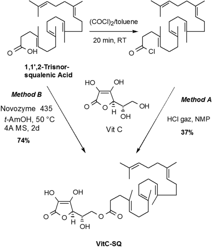
        Vitamin C (ascorbic acid) is a charged and hydrophilic natural antioxidant with skin antiaging and photoprotective effects. Because it penetrates poorly into the skin, lipidic derivatives more able to enter stratum corneum have been designed as antioxidant excipient for topical formulations. Many fatty acids esterified forms of the C-6 primary hydroxyl group of vitamin C were thus introduced in pharmaceutical or cosmetic compositions. For example, 6-O-palmitoyl-L-ascorbic acid was shown to reduce the appearance of wrinkles, cracks, crevices and puffiness around the eyes23 and 6-O-stearoyl-L-ascorbic acid was found to retard skin aging24 and to be a potent inhibitor of hyaluronidase enzymes25. Many other derivatives including esters of oleic acid26, linoleic27, myristic acid28, retinoic acid29, docosahexaenoic acid30, etc. have been evaluated for potential application in cosmetic and as antioxidant in food industry. The anchoring of the lipidic moiety on the lateral chain of ascorbic acid preserves the redox properties of the tetronic core. Furthermore, this design considerably simplifies the synthesis since the primary hydroxyl group is the most nucleophilic function. We thus envisaged to access 6-squalenoyl ascorbic acid (Vit C–SQ) by direct condensation of 1,1′,2-trisnor-squalenic acid, easily available from squalene31,32, without protection of the tetronic ring. Several synthesis strategies were described to obtained SQ derivatives33,34,35. However, in the case studied here, most conventional methods failed to give the expected material. We finally found that when ascorbic acid was treated with squalenyl chloride in HCl saturated N-methyl-pyrrolidinone (NMP) according to the method of Yazawa et al.36, the desired 6-O-squalenoyl-L-ascorbic acid could be obtained in 30–35% yield after chromatographic purification. Although this method had the merit of giving the desired product, it suffered from extensive degradation during the removal of the high boiling solvent NMP. To bypass this hurdle, we examined the enzymatic acylation of ascorbic acid with stabilized lipase. Biocatalysts offered mild conditions and improved regioselectivity for the acylation of ascorbic acid, particularly when sensitive unsaturated fatty acids were involved37,38,39. The 6-O-squalenoyl-L-ascorbic acid was thus synthetized by treatment of a 2:1 mixture of ascorbic acid and squalenic acid with Novozyme 435, a lipase acrylic resin from Candida antarctica in t-AmOH at 50 °C for 2 days, using molecular sieves as drying agent. In these conditions the Vit C–SQ was obtained in 74% yield as a thick oil after chromatographic purification (Fig. 1). The Vit C–SQ derivative was stable upon 2 years storage in a freezer at − 20 °C.

        Figure 1
        figure1

        Synthesis of Vit C–SQ through chemical esterification (Method A) or enzymatic acylation (Method B).

        Full size image

        Formulation of Vit C–SQ

        Vit C–SQ and Vit C–Palmitate (used as a control) were formulated as creams to be directly applied on human explants. These oily compounds were dispersed in SQ at equivalent concentrations of Vit C (3 or 5 wt%) and homogenized by magnetic stirring. The Vit C–SQ and Vit C–Palmitate formulations were viscous, with an oily texture and slightly yellow color. Free Vit C could not be dispersed in SQ, so it was incorporated in carboxymethyl cellulose at equivalent contents.

        Skin morphology was improved by Vit C–SQ bioconjugate

        Formulations containing free or conjugated Vit C or SQ alone, were applied repeatedly for 10 days upon stratum corneum of human skin explants collected from two 30-year females and maintained in a proprietary survival medium. Untreated control explants from each donor were kept in the same conditions for the same time. The explants from both donors received distinct treatment panels, i.e. donor 1 received for 10 days Vit C either free or coupled to SQ or to Palmitate, at 1 or 3  wt% final Vit C concentration, while donor 2 received for 10 days free Vit C 5 wt% or Vit C–SQ at 3 or 5 wt%, or SQ alone. Thus, the experiment with Vit C–SQ 3 wt% was repeated in both donors, allowing comparison.

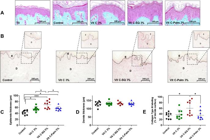
        In fixed and colored tissue sections from each explant, the skin morphology as well as the epidermis and dermis thickness were investigated. A significant increase of epidermis thickness was observed with Vit C–SQ 3 wt% in donor 1 (Fig. 2A, C), i.e. + 60% compared to the untreated control (69 ± 12 µm versus 43 ± 7 µm, respectively; p < 0.001). The thickening effect was more limited with the same concentration of free Vit C (+ 28%; p < 0.05) or of Vit C–Palmitate (+ 31%; p < 0.05). Consistently, in donor 2 a 22% increase in epidermis thickness was observed with Vit C–SQ 3 wt% (p < 0.05), as well as, with Vit C–SQ 5 wt% (p < 0.05), while only a non significant 14% increase was observed with free Vit C 5 wt% and no variation with SQ alone (Supplementary Fig. S1A, C), thus confirming the beneficial effect of Vit C–SQ on skin thickness. Epidermal thickness before treatment (40 ± 4 µm) was not significantly different from the value after 10 days without treatment (43 ± 7 µm; T0) in donor 1 (Fig. 2A). On the other hand, dermis thickness (128 ± 14 µm in control from donor 1; 125 ± 4 µm in control from donor 2) was not modified whatever the Vit C formulation used in both donors nor with SQ in donor 2 (Fig. 2D, Supplementary Fig. S1D). Finally, of all Vit C formulations studied, Vit C–SQ resulted in the highest gain of epidermis thickness.

        Figure 2
        figure2

        Topical treatment of human skin explants (donor 1) for 10 days with three formulations containing 3 wt% Vit C, increased epidermis thickness and collagen type III labeling in dermis. (A) In fixed and stained (Masson trichrome) whole skin sections, epidermis (pink) and dermis (blue) layers were observed before (T0) and after the treatment period. The control explant was kept untreated for 10 days. (B) Fixed skin sections labeled with goat anti-human collagen III antibody and revealed by DAB are shown. Pictures were realized with a tri CCD DXC 390P camera (Sony) and stored with Leica IM1000 software, version 1.10 (www.leica-microsystems.com). (C) The epidermis thickness and (D) dermis thickness were measured in each explant (n = 7 to 9 images; mean of 3 measures per image). (E) The intensity of collagen III immuno-staining signal in dermis was measured as the percentage of analyzed surface (n = 9 images; mean of 3 measures per image). One-way ANOVA with Tukey's post-test (***p < 0.001, *p < 0.05) was used to compare treatments. Graphs were done with GraphPad Prism version 5.0.0 for Windows (GraphPad Software, San Diego, California USA, www.graphpad.com). Scale bar = 100 µm (panel A), and 200 μm (panel B).

        Full size image

        The skin biological pathways were studied at molecular level

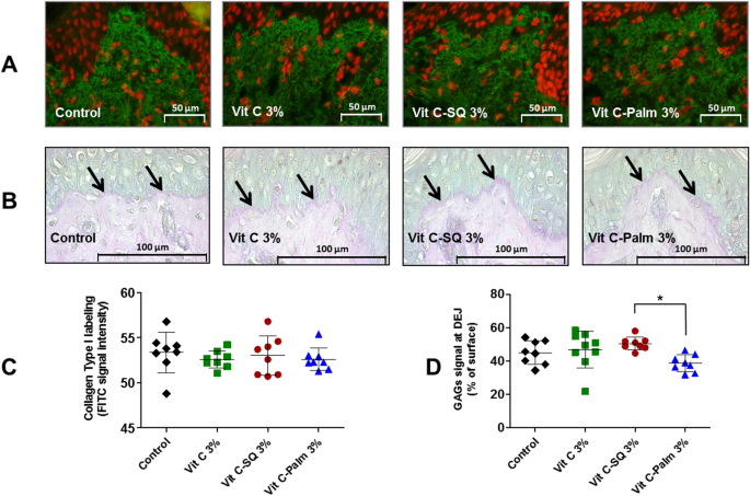
        The expression of proteic (collagens; Figs. 2B, 3A) as well as glucidic skin markers (GAGs; Fig. 3B, Supplementary Fig. S1B, E) has been measured by immuno-histochemistry in human tissue explants after the different topical treatments.

        Figure 3
        figure3

        Effects of ex-vivo topical treatment of human skin explants (donor 1) for 10 days with three formulations containing 3 wt% Vit C, upon collagen type I expression in whole skin and GAGs staining at dermal–epidermal junction (DEJ). (A) Collagen type I was labeled by a rabbit anti-human collagen I antibody with biotin/streptavidin amplification and FITC fluorescence revelation (green) and cell nuclei stained with propidium iodide (red). Pictures were realized with a tri CCD DXC 390P camera (Sony) and stored with Leica IM1000 software, version 1.10 (www.leica-microsystems.com). (B) Staining of fixed skin sections with Schiff reagent revealed the neutral GAGs as purple pink near DEJ (arrows), at the bottom of upper epidermis layer. (C) One-way ANOVA with Tukey's post-test was used to compare collagen I signal (n = 8 images; mean of 3 measures per image), relative to untreated control (not significant). (D) GAGs coloration measured as percent of DEJ surface (n = 8 to 9 images; mean of 3 measures per image) from each explant was compared in formulations and untreated control by one-way ANOVA with Tukey's post-test (*p < 0.05). Graphs were done with GraphPad Prism version 5.0.0 for Windows (GraphPad Software, San Diego, California USA, www.graphpad.com). Scale bar = 50 µm panel A, and 100 μm panel C.

        Full size image

        To gain insights on the molecular pathways behind the observed effects of Vit C–SQ compared to Vit C–Palmitate, to free Vit C or to SQ, a set of 15 genes of interest were selected according to their known relationships with skin physiopathology, which have been documented using the Ingenuity Pathway Analysis (IPA) software40 (Supplementary Fig. S2). The expression of these genes was measured by RT-qPCR in the same skin explants previously treated with the various formulations of Vit C or SQ alone (Supplementary Fig. S3). The expression of some of these genes was also evaluated in epidermal and dermal compartments previously separated by laser capture microdissection. For some genes, the transcriptional effects were higher upon epidermis compared to dermis, which might be related to the epidermis thickening described before (Supplementary Fig. S4).

        It can be concluded from this study that the Vit C–SQ complex has modified in the strongest extent the transcriptional expression of most of the target genes studied, in a globally reproducible manner for the Vit C–SQ 3 wt% tested in two independent donors. The effects observed in whole skin were also consistent with the sum of the effects measured in separately microdissected epidermis and dermis in the two donors. Moreover, the transcriptional expression data showed consistency with histological data. Hereafter the effects of this bioconjugate upon the various biological functions explored will be reviewed.

        Vit C–SQ favors biosynthesis and maintenance of structural fibers of the skin

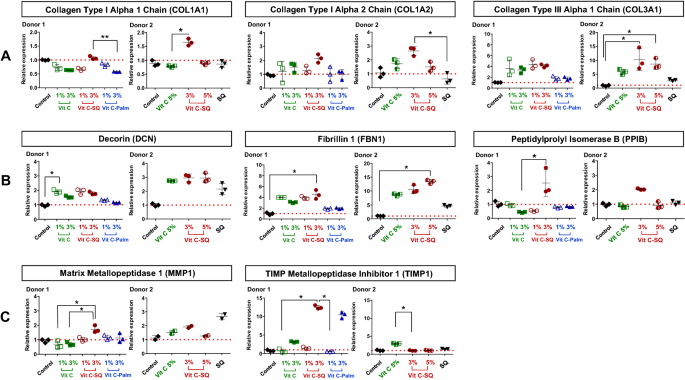
        Collagen is a major constituent of skin where it is structured into fibers representing the major part of dermis mass17. Type III collagen is a homotrimer forming a right-handed triple helix4 which acts as a major structural constituent of the extracellular matrix. The immuno-histochemistry studies performed in donor 1 showed that the collagen III protein content was twice higher in papillary dermis after 10-days treatment with 3 wt% Vit C–SQ formulation comparatively to untreated control, i.e. collagen III signal represented 60 ± 23% versus 34 ± 10% of dermis surface, respectively (p < 0.05). The more limited effects of Vit C–Palmitate (+ 4%) and of free Vit C (+11%) were not significant (Fig. 2B, E). Consistently, this stimulating effect of collagen III biosynthesis by Vit C–SQ upon was also observed in both donors at gene expression level. The COL3A1 mRNA transcript was increased in donor 1 (fourfold) and in donor 2 (11-fold; p < 0.05) after Vit C–SQ 3 wt% treatment versus control, and similarly with Vit C–SQ 5 wt% in donor 2 (tenfold; p < 0.05) (Fig. 4A). Comparatively, the free Vit C produced a smaller and not significant increase in COL3A1 expression in both donors, while Vit C–Palmitate was almost devoid of any effect in donor 1, as well as SQ alone in donor 2.

        Figure 4
        figure4

        Vitamin C formulations influenced the expression in human skin explants, of the mRNA transcripts of (A) genes encoding collagens type I and type III, (B) genes controlling skin fibrils assembly and (C) genes regulating the extracellular matrix (ECM). Fold changes of each transcript were calculated as 2−ΔCq from Cq normalized by NORMA-gene algorithm56, in whole skin explants treated with 1–3 wt% free or formulated Vit C (in donor 1) or with 3–5 wt% free or squalenized Vit C or with squalene (SQ) alone (in donor 2), relative to the untreated control (set to 1, red dotted line). The data from one of two or three analyzed explants are presented (n = 3 per group). Kruskal–Wallis non-parametric test followed by Dunn's post-test was used to determine significance between the groups (**p < 0.01, *p < 0.05). Graphs were done with GraphPad Prism version 5.0.0 for Windows (GraphPad Software, San Diego, California USA, www.graphpad.com).

        Full size image

        The collagen I protein content was not modified in dermis from skin explants treated for 10 days with any of the 3 wt% Vit C formulations tested, according to immuno-histochemistry results in donor 1 (Fig. 3A, C). Among the genes encoding type I procollagen41, the expression of COL1A1 gene, encoding collagen type I alpha 1 chain, was not modified by Vit C–SQ in donor 1 and was poorly raised in donor 2 (1.6-fold, p < 0.05). The COL1A2 gene encoding collagen type I alpha 2 chain was slightly overexpressed after Vit C–SQ 3 wt% treatment in both subjects studied, i.e. 2.1 to 2.6-fold (p < 0.05). In contrast, free Vit C, Vit C–Palmitate or SQ alone had no significant effect in collagens I gene expression (Fig. 4A).

        Our transcriptional data obtained from skin layers after microdissection suggest that the overexpression of collagens I and III induced by Vit C–SQ could take place in dermis (Supplementary Fig. S4), consistently with described stimulation of collagen production by Vit C in dermis fibroblasts17. Overall, among the collagen-encoding genes studied here, COL3A1 was the most strongly modified and Vit C–SQ was the most active formulation since it stimulated 2–4 times more its expression than that of COL1A2. These results are in line with studies on liposomal Vit C showing increased production of collagen in pig ear skin35. Our study demonstrates that collagen production is stimulated by Vit C derivatives also in human skin. Finally, the observed effects of Vit C–SQ upon collagens I and III contribute to a limited I / III ratio, which is typical of young skins11,12. This could characterize an anti-aging action for this bioconjugate.

        Decorin (DCN)42,43 and fibrillin 1 (FBN1)44 are involved in the assembly and maintenance of collagen fibrils and elastic microfibrils, thus contributing to skin strength and suppleness. The corresponding genes were significantly overexpressed in whole skin explants from both subjects, treated with Vit C formulations, with a higher effect of the Vit C–SQ conjugate, i.e. DCN twofold to threefold and FBN1 fourfold to tenfold (p < 0.05), while other formulations had lower or no effect (Fig. 4B). In microdissected skin layers, the transcriptional effect of Vit C–SQ was observed in epidermis for FBN1, thus this might be related to the epidermis thickening described above; however, it occurred at a similar extent in dermis and epidermis for DCN (Supplementary Fig. S4). Moreover, the gene encoding peptidylprolyl isomerase B (PPIB) which contributes to collagen binding, accelerates proteic folding and favors tensile strength of skin45, was twice overexpressed with Vit C–SQ 3 wt% (p < 0.05), while it was not affected by free Vit C or Vit C–Palmitate 3 wt% or by SQ (Fig. 4B).

        Vit C–SQ positively regulates the components of ECM

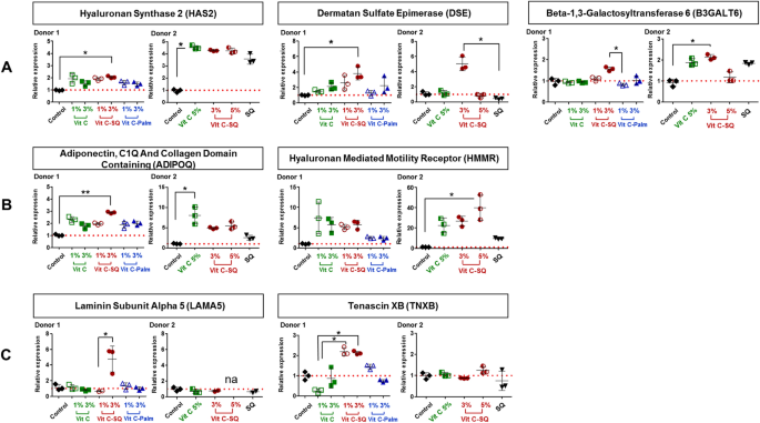
        The matrix metalloproteinases (MMPs) are known to contribute to basement membrane and ECM degradation through the fragmentation of collagens, during skin aging and ultraviolet irradiation46. In our study, the Vit C–SQ complex stimulated 6-times more in donor 1 the transcriptional expression of TIMP metallopeptidase inhibitor of MMPs (TIMP1; 12-fold, p < 0.05; Fig. 4C) and twice more the expression of COL3A1 transcript (fourfold; Fig. 4A), than the expression of MMP1 transcript (1.7-fold, p < 0.05; Fig. 4C) which encodes the collagenase involved in the degradation of type I and III collagens41. Such effects can contribute to preserve the existing collagen. This is consistent with the observed favorable action of Vit C–SQ upon the production of collagens I (Fig. 4A) and III (Figs. 2B, E, 4A) and on the expression of decorin (Fig. 4B), which locates at the surface of collagen fibers thus protecting them against cleavage by MMPs43. Moreover, members of two families of ECM glycoproteins, i.e. laminins, which are the major non collagenous constituent of basement membranes, and tenascins, involved in wound healing47, were also positively modulated at gene level in donor 1 by Vit C–SQ 3 wt% only: laminin subunit alpha 5 (LAMA5; fourfold, p < 0.05) and tenascin XB (TNXB; twofold, p < 0.05) (Fig. 5C).

        Figure 5
        figure5

        Vitamin C formulations influenced the expression in human skin explants, of the mRNA transcripts of (A) genes encoding enzymes of GAGs biosynthesis, (B) genes controlling cell growth and motility, and (C) genes regulating ECM. Fold changes of each transcript were calculated as 2−ΔCq from Cq normalized by NORMA-Gene algorithm56, in whole skin explants treated with 1–3 wt% free or formulated Vit C (in donor 1) or with 3–5 wt% free or squalenized Vit C or with squalene (SQ) alone (in donor 2), relative to the untreated control (set to 1, red dotted line). The data from one of two or three analyzed explants are presented (n = 3 per group). Kruskal–Wallis non-parametric test followed by Dunn's post-test was used to determine significance between the groups (**p < 0.01, *p < 0.05). Graphs were done with GraphPad Prism version 5.0.0 for Windows (GraphPad Software, San Diego, California USA, www.graphpad.com).

        Full size image

        Vit C–SQ is favorable to GAGs content in skin

        Hyaluronic acid (hyaluronan, HA) is an acidic GAG predominantly present in skin where it retains water and promotes cell motility, adhesion, proliferation and tissue organization. Moreover, HA accumulates in ECM in early stages of wound healing48. Hyaluronan synthase 2 (HAS2) mediates the biosynthesis of HA49 and is described as its main synthase in skin50. The biosynthesis of HA could be stimulated by Vit C–SQ 3 wt% in skin, as suggested by the overexpression in whole skin explants, of the HAS2 gene, i.e. twofold (p < 0.05) in donor 1 and fourfold in donor 2 (Fig. 5A).

        This is also consistent with the observed overexpression to a similar extent after treatment with Vit C–SQ 3 wt% (threefold, p < 0.01 in donor 1), of the gene encoding adiponectin (ADIPOQ), which promotes biosynthesis of HA along with HAS2 transcript51 (Fig. 5B). The effect of Vit C–SQ upon this gene supports the observed thickening of the epidermis (Fig. 2A, C), since adiponectin favors cell growth and tissue remodeling through the binding and sequestration of growth factors. The hyaluronan mediated motility receptor (HMMR), which is a major HA receptor and regulates cell growth, motility and contact inhibition48 was also strongly overexpressed (sixfold in donor 1; 40-fold, p < 0.05 in donor 2) by Vit C–SQ 3 or 5 wt% in whole skin (Fig. 5B).

        Moreover, histochemistry staining of neutral GAGs, which act as growth factors reservoirs at the dermal–epidermal junction (DEJ)48, showed a stronger signal at DEJ in skin explants treated with Vit C–SQ 3 wt% compared to Vit C–Palmitate 3 wt% in donor 1 (+ 30%, p < 0.05; Fig. 3B, D). Consistently, skin explants treated with Vit C–SQ 3 wt% exhibited a 50%-higher GAGs signal at DEJ, compared to free Vit C 5 wt% in donor 2 (p < 0.05; Supplementary Fig. S1B, E). The genes encoding two enzymes involved in GAGs biosynthesis were consistently overexpressed in both donors after Vit C–SQ 3 wt% treatment, i.e. beta-1,3-galactosyltransferase 652 (B3GALT6; twofold, p < 0.05) and dermatan sulfate epimerase53 (DSE; fourfold, p < 0.05) (Fig. 5A).

        Conclusion

        Topical application of Vit C on the skin with a strategy for the delivery through the SC has proved its usefulness as a complement to Vit C nutritional intake, as well as, in various cases of skin damage situations, such as UV irradiation, inflammation, redox unbalance, aging and wound healing4,19. At equivalent concentrations in Vit C, the Vit C–SQ bioconjugate exerted higher beneficial actions on human skin comparatively to free Vit C or Vit C linked to palmitic acid. Not only Vit C–SQ triggered a marked epidermis thickening, but it also induced the production of collagen III typical of young skins, as well as, the assembly of collagen fibers and elastic microfibrils and favored cutaneous hydration by stimulating production of mucopolysaccharides which have high water retention ability. With further studies, Vit C–SQ based formulations could find potential applications in cosmetics and dermatology care. In a nutshell, the VitC-SQ bioconjugate demonstrated a beneficial action on skin, although its mechanism of action still remains uncovered. It is, however, hypothesized that, apart the preservation of the Vit C anti-oxidant activity through the conjugation to SQ, this lipid may also favor skin penetration and possible interaction with cell membranes.

        Experimental section

        Synthesis of Vit C–SQ

        IR spectra were obtained as solid or neat liquid on a Fourier Transform Bruker Vector 22 spectrometer. Only significant absorptions are listed. The 1H and 13C NMR spectra were recorded on Bruker Avance 300 (300 MHz and 75 MHz, for 1H and 13C, respectively) or Bruker Avance 400 (400 MHz and 100 MHz, for 1H and 13C, respectively) spectrometers. Recognition of methyl, methylene, methine, and quaternary carbon nuclei in 13C NMR spectra rests on the J-modulated spin-echo sequence. Mass spectra were recorded on a Bruker Esquire-LC. Analytical thin-layer chromatography was performed on Merck silica gel 60F254 glass precoated plates (0.25 mm layer). Column chromatography was performed on Merck silica gel 60 (230–400 mesh ASTM). Toluene and N-methyl pyrrolidone (NMP) were distilled from calcium hydride, under a nitrogen atmosphere. t-AmOH, was dried of sodium and distilled. All reactions involving air- or water-sensitive compounds were routinely conducted in glassware which was flame-dried under a positive pressure of nitrogen or argon. Ascorbic acid, oxalyl chloride, squalene, NMP and Novozyme 435 (L4777) were purchased from Sigma-Aldrich Chemical Co., France. Chemicals obtained from commercial suppliers were used without further purification. 1,1′,2-tris-norsqualenic acid was prepare according to Ceruti et al.32. For comparison purposes, palmitoyl ascorbate was prepared from palmitic acid and ascorbic acid according to litterature54.

        4,8,13,17,21-Pentamethyl-docosa-4,8,12,16,20-pentaenoyl chloride (2)

        A solution of trisnorsqualenic acid (1.80, 4.5 mmol) in anhydrous toluene (10 mL) was degassed by bubbling a stream of nitrogen through the solution for 5 min. Oxalyl chloride (1.74 g, 13.8 mmol) was added dropwise at 20 °C. The reaction mixture was stirred for 3 h at the same temperature and concentrated under reduced pressure to give the title compound as a yellow oil which is used directly in the next step. IR (film) ν: 2920, 1799, 1443, 1382, 955, 893 cm−1; RMN 1H (300 MHz, CDCl3) δ : 5.23–5.10 (m, 5 H), 2.45 (t, J = 7.3 Hz, 2 H), 2.30 (t, J = 7.5 Hz, 2H), 2.15–1.95 (m, 16 H), 1.70 (s, 3H), 1.62 (s, 15H) ppm; RMN 13C (75 MHz, CDCl3), δ :173.2 (C, COCl), 135.1 (C, C=CH), 134.8 (C, C=CH), 134.6 (C, C=CH), 131.4 (C, C=CH), 131.1 (C, C=CH), 126.6 (CH, C=CH), 124.7 (CH, C=CH), 124.4 (CH, C=CH), 124.3 (2CH, C=CH), 45.8 (CH2), 39.7 (2CH2), 34.4 (CH2), 34.6 (CH2), 28.2 (2CH2), 26.8 (CH2), 26.7 (CH2), 26.5 (CH2), 25.7 (CH3, C=C(CH3)2), 17.6 (CH3), 16.0 (CH3), 15.9 (2 CH3), 15.8 (CH3) ppm.

        6-O-[4,8,13,17,21-Pentamethyl-docosa-4,8,12,16,20-pentaenoyl] ascorbic acid, method A

        A stream of dry HCl was passed through 5 mL of N-methylpiperidone in a washing bottle until the entire mass solidified. N-methylpyrrolidinone was added to get a clear solution (~ 5 mL) which was transferred into a 50 mL round bottom flask. Ascorbic acid (0.7 g, 4.0 mmol) was added and the mixture was stirred until complete dissolution. The reaction mixture was cooled to 0 °C and squalenoyl chloride (418 mg, 1.0 mmol) was added. After being stirred for 20 h at room temperature, water was added (20 mL) and the mixture was extracted with ethyl acetate (4 × 20 mL). The combined organic layers were washed with water (2 × 5 mL) dried over MgSO4 and concentrated under reduced pressure. The residual NMP was removed under reduced pressure using dry ice rotavapor (60 °C, 0.05 mm Hg,) to leave an oil which was purified by chromatography over silica gel eluting with ethyl acetate and then AcOEt/MeOH (98:2) to give the title compound as a pale yellow thick oil (190 mg, 34%).

        6-O-[4,8,13,17,21-Pentamethyl-docosa-4,8,12,16,20-pentaenoyl] ascorbic acid, method B

        To a suspension of ascorbic acid (200 mg, 2 mmol) in anhydrous tert-amyl alcohol (5 mL) was successively added Novozyme 435 (150 mg), tris-norsqualenic acid (200 mg, 1.0 mmol) and 1 g of 4 Å molecular sieves. The flask was fixed to a rotavapor and slowly stirred at 50 °C (water bath). After 48 h, the reaction mixture was cooled and filtered through a plug of Celite. The solid was thoroughly washed with AcOEt and the filtrate was concentrated under reduced pressure. The crude product was purified by chromatography over silica gel eluting with ethyl acetate and then AcOEt/MeOH (98:2) to give the title compound as a pale yellow thick oil (209 mg, 74%); [α]D =  + 10 (c = 0.5, EtOH); IR (film) ν: 3600–2800, 2976, 2857, 1744, 1696, 1443, 1382, 1350, 1296, 1151, 1119, 1043, 983, 844 cm−1; NMR 1H (360 MHz, MeOH-d4) δ: 5.20–5.05 (m, 5H), 4.72 (d, J = 2.0 Hz, 1H, H-4), 4.24 (dd, J = 11.16 Hz , J = 6.84 Hz, 1H, H-6), 4.18 (dd, J = 11.16 Hz , J = 6.12 Hz, 1H, H-6), 4.08 (ddd, J = 6.84 Hz, J = 6.12 Hz, J = 2.0 Hz, 1H, H-5), 2.46 (t, J = 7.74 Hz, 2H, COCH 2CH2), 2.30 (t, J = 7.56 Hz, 2H, COCH2CH 2), 2.12–1.92 (m, 16H), 1.63 (d, J = 1.08 Hz, 3H, HC = C(CH 3)2), 1.62 (s, 3H), 1.60 (s, 12H) ppm; NMR 13C (90.5 MHz, CDCl3) δ: 174.6 (C, CO), 173.1 (C, C1), 154.0 (C, C3), 136.0 (C, C=CH), 135.9 (C, C=CH), 135.8 (C, C=CH), 134.4 (C, C=CH), 132.0 (C, C=CH), 126.3 (CH, C=CH), 125.6 (CH, C=CH), 125.5 (CH, C=CH), 125.4 (2CH, C=CH), 120.1 (C, C2), 77.1 (CH, C4), 68.0 (CH, C5), 65.7 (CH2, C6), 40.9 (CH2), 40.8 (CH2), 40.7 (CH2), 35.7 (CH2), 33.9 (CH2), 29.2 (2CH2), 27.8 (CH2), 27.7 (CH2), 27.6 (CH2), 25.9 (CH3, C=C(CH3)2), 17.8 (CH3), 16.2 (2CH3), 16.1 (CH3) , 16.0 (CH3) ppm; SM (+APCI) m/z (%): 559.6 (100) [M + H]. The IR, 1H and 13C NMR spectra are presented in Supplementary Figs. S5, S6 and S7, respectively.

        Preparation of the Vit C–SQ cream

        Vit C–SQ (30.1 mg, 0.054 mmol) was weighted in a 5 mL vial. SQ (222 mg, 250 mL, 0.54 mmol) was added via syringe and the mixture was vortexed for 2 min. The obtained mixture was then sonicated using an ultrasonic cleaning bath to give a homogenous cream containing 3 mmol% of Vit C. A similar procedure was followed to obtain the Vit C–SQ cream containing 5 mmol% of Vit C.

        Formulations for ex vivo skin testing

        Explants from two donors aged 30 years were treated with the following formulations: (1) Vit C at 1 or 3 wt% in an aqueous cream of carboxymethyl cellulose (Sigma Aldrich) (2) Vit C–SQ conjugate dispersed in SQ; (3) Vit C–Palmitate complex; (4) squalenic acid in SQ (70:30, w:w) and (5) pure SQ. Final concentrations of Vit C in the various formulations were 1, 3 or 5 wt%.

        Ex vivo activity testing on human skin

        BIO-EC Laboratory possesses an authorization from the Bioethics group of the General Director Services of the French Research and Innovation Ministry (registered under n°DC-2008-542) to use human skin from surgical waste since 5th May 2010. The study was performed in accordance with the Declaration of Helsinki after the patients had given informed consent to use their skin samples by BIO-EC Laboratory.

        Skin explants (1-cm diameter) were recovered from an esthetic abdominal surgery in two 30-years old women and maintained in vitro for 9- or 10-days survival at 37 °C, in wet atmosphere enriched with 5% CO2, in BIO-EC's Explants Medium (BEM, BIO-EC Laboratory). At days 0, 1, 3, 6 and 8, explants received topical treatments with each cosmetic formulation (2 mg per explant) spread with a spatula, i.e. free Vit C, Vit C–SQ or Vit C–Palmitate at 1%, 3% or 5% (w:w). As a reference, pure SQ was also tested in one donor (donor 2). Untreated explants sampled at day 10 for donor 1 or 2 showed identical morphology as day-0 explants and were used as controls. Three explant replicates were prepared for each biological condition. Each skin explant was divided in two parts, one was fixed with ordinary Bouin and paraffin-embedded, the other one was frozen at − 80 °C until microdissection and genomic studies were performed.

        Histological and immunohistochemistry studies

        Five-5 µm tissue sections were prepared using a Minot RM 2125 microtome (Leica) and sticked on superfrost silanized glass slides. General tissue morphology was observed after Goldner-modified Masson trichrome staining. Glycosaminoglycans (GAGs) were examined on paraffin-embedded skin sections after staining with Schiff reagent. Neutral GAGs which link growth factors appear as purple pink near the DEJ. Optical microscopy observations were done with an Orthoplan microscope (Leica) at X40 magnification. Pictures were obtained with a tri CCD DXC 390P camera (Sony) and stored with Leica IM1000 software, version 1.10 (Leica). Collagen I immunostaining was done on frozen sections with 800-fold diluted polyclonal rabbit anti-human collagen I antibody (Monosan ref PS047) for one hour at room temperature, with a biotin/streptavidin amplification system, fluorescence revelation (FITC Caltag SA 1001) and nuclei staining with propidium iodide. Collagen III immunostaining was done on formol-fixed and paraffin-embedded slices with 50-fold diluted polyclonal goat anti-human collagen III antibody (SBA ref 1330-01), overnight at room temperature, with a Vectastain RTU Universal VECTOR avidin/biotin amplification system, DAB revelation.

        Laser capture microdissection

        Some skin explants were microdissected in order to separate dermis from epidermis. Serial sections (12-µm-thick human skin frozen tissue specimens) were placed on membrane slides (Carl Zeiss MicroImaging). Cresyl violet staining was performed by using a protocol from Zeiss Labs. Epidermal and dermal tissues were selectively dissected using the PALM MicroBeam system (Carl Zeiss). The dissection procedure was validated by measuring the differential expression of transcripts from genes specifically expressed in each tissue.

        RNA extraction

        Whole skin samples of 40–100 mg weight were homogenized in 1 mL TRI-Reagent (Euromedex), by agitation in a Precellys grinder with MK28-R metallic beads (Bertin) for two cycles of 3 × 20 s at 6500 rpm. After precipitation in 500 µL of isopropanol, RNA was high-speed centrifugated and the RNA pellet was washed twice with 75% ethanol, then solubilized in 30 µL of RNase-free water and stored at − 80 °C until molecular analyses. RNA purity and quantity were assessed by UV absorbance readings and RNA integrity was evaluated by capillary electrophoresis using RNA 6000 Nano chips and the 2100 Bioanalyzer (Agilent Technologies). RNA from microdissection samples were extracted in 300 µL TRI-Reagent, precipitated in 500 µL isopropanol overnight at − 20 °C, solubilized in 20 µL of RNase-free water and quality checked using Total RNA 6000 Pico chips and the 2100 Bioanalyzer. All RNA extracts showed homogeneous quality levels.

        Amplification and reverse transcription of RNA

        Prior to RT-qPCR study, 1 µg of total RNA from whole skin was amplified with the Quick Amp Labeling Kit (Agilent Technologies, ref 5190-0444), consisting of a double strand cDNA synthesis with MMLV-RT and oligo-dT-T7 promoter priming, followed by in vitro transcription with T7 RNA polymerase. Ten microliters of each amplified cRNA was directly reverse transcribed in 20-µL reactions, using Transcriptor First Strand cDNA Synthesis Kit (Roche Diagnostics) and gene-specific priming with a 3-µL mix of qPCR primers designed for all genes studied (0.5 µM of each primer as final concentration in RT). The RNA-primers mix was incubated for 5 min at 65 °C, cooled on ice, completed to 20 µL with RT reagents and incubated for 30 min at 55 °C before enzyme inactivation for 5 min à 85 °C. Similarly, amplification of RNA extracted from each microdissected skin layer was performed from 10 µL of purified RNA solution and then RT was done with a priming mix at 0.07 µM final concentration in RT.

        Real-time quantitative PCR

        PCR primers obtained from the qPrimerDepot database55 or designed with Primer Designing Tool (https://www.ncbi.nlm.nih.gov/tools/primer-blast/) were chosen overlapping an intron or hybridizing an exon junction when possible and checked for in silico specificity before synthesis by Eurofins Operon (Supplementary Table S1). qPCR analysis was done in triplicate 15-µL reactions with SYBR Premix Ex Taq Tli RNase H Plus mix (Takara) including 5 µL of 30-fold diluted cDNA and 0.5 µM of each primer. A 2-steps PCR program with hybridization at 61 °C followed by specificity check on fusion curves, was performed on CFX384 thermal cycler (Bio-Rad Laboratories). Quantitative values were normalized using the NORMA-Gene method56 for whole-skin data and with GAPDH and ACTB housekeeping genes for microdissection data57. Relative expression ratios were calculated by the delta-delta Cq method58, based on a theoretical efficiency considered at 100%, using as calibrator the untreated condition of similar duration as treated samples (day 10).

        Statistical analysis

        Data analysis was performed with GraphPad Prism version 5.0.0 for Windows (GraphPad Software, San Diego, California USA, www.graphpad.com). For epidermis and dermis thickness, collagen type I labelling and GAGs staining at DEJ, differences between treatments were tested using one-way ANOVA with Tukey's multiple comparison post-test, with variances homogeneity checking by Bartlett's test. For collagen type III labelling and gene expression fold-changes in whole skin explants, differences between treatments were tested using Kruskal–Wallis and post hoc Dunn's test. For expression data from microdissected skin, differences were tested by two-way ANOVA followed by Bonferroni's multiple comparison test. Differences between conditions were considered significant when p values < 0.05 (*p < 0.05; **p < 0.01; ***p < 0.001). Quantitative data were graphically presented as mean ± standard deviation.

        Data availability

        The datasets generated and/or analyzed during the current study are provided as Supplementary Data.

        References

        1. 1.

          Darr, D., Combs, S., Dunston, S., Manning, T. & Pinnell, S. Topical vitamin C protects porcine skin from ultraviolet radiation-induced damage. Br. J. Dermatol. 127, 247–253 (1992).

          CAS  PubMed  Article  PubMed Central  Google Scholar

        2. 2.

          Austria, R., Semenzato, A. & Bettero, A. Stability of vitamin C derivatives in solution and topical formulations. J. Pharm. Biomed. Anal. 15, 795–801 (1997).

          CAS  PubMed  Article  PubMed Central  Google Scholar

        3. 3.

          Wang, K. et al. Role of vitamin C in skin diseases. Front. Physiol. 9, 819 (2018).

          PubMed  PubMed Central  Article  Google Scholar

        4. 4.

          Ravetti, S. et al. Ascorbic acid in skin health. Cosmetics 6, 58–69 (2019).

          CAS  Article  Google Scholar

        5. 5.

          Matsuda, S., Shibayama, H., Hisama, M., Ohtsuki, M. & Iwaki, M. Inhibitory effects of novel ascorbic derivative VCP-IS-2Na on melanogenesis. Chem. Pharm. Bull. 56, 292–297 (2008).

          CAS  PubMed  Article  PubMed Central  Google Scholar

        6. 6.

          Traikovich, S. S. Use of topical ascorbic acid and its effects on photo-damaged skin topography. Arch.. Otorhinol. Head Neck Surg. 125, 1091–1098 (1999).

          CAS  Article  Google Scholar

        7. 7.

          Talakoub, L., Neuhaus, I. M. & Yu, S. S. Cosmeceuticals. In Cosmetic Dermatology. Requisites in Dermatology 1st edn, Vol. 1 (eds Alam, M. et al.) 13–14 (Saunders Elsevier, Gurgaon, 2009).

          Google Scholar

        8. 8.

          Rabat Sarpooshi, H., Mortazavi, F., Mojtaba, V. & Tabarayee, Y. The effects of topical vitamin C solution on burn wounds granulation: a randomized clinical trial. J. Biomed. 1(4), e8301 (2016).

          Article  Google Scholar

        9. 9.

          Boyce, S. T., Supp, A. P., Swope, V. B. & Warden, G. D. Vitamin C regulates keratinocyte viability, epidermal barrier, and basement membrane in vitro, and reduces wound contraction after grafting of cultured skin substitutes. J. Investig. Dermatol. 118, 565–572 (2002).

          CAS  PubMed  Article  PubMed Central  Google Scholar

        10. 10.

          Nielsen, M. J. & Karsdal M. A. Biochemistry of collagens, laminins and elastin. structure, function and biomarkers (ed. Karsdal, M. A.) 21–30 (Elsevier, 2016).

        11. 11.

          Cheng, W., Yan-hua, R., Fang-gang, N. & Guo-an, Z. The content and ratio of type I and III collagen in skin differ with age and injury. Afr. J. Biotechnol. 10, 2524–2529 (2011).

          Google Scholar

        12. 12.

          Rong, Y. H., Zhang, G. A., Wang, C. & Ning, F. G. Quantification of type I and III collagen content in normal human skin in different age groups. Zhonghua Shao Shang Za Zhi 24, 51–53 (2008).

          PubMed  PubMed Central  Google Scholar

        13. 13.

          Liu, X., Wu, H., Byrne, M., Krane, S. & Jaenisch, R. Type III collagen is crucial for collagen I fibrillogenesis and for normal cardiovascular development. PNAS 94, 1852–1856 (1997).

          ADS  CAS  PubMed  Article  PubMed Central  Google Scholar

        14. 14.

          Lee, A. C. & Tojo, K. Characterization of skin permeation of vitamin C: theoretical analysis of penetration profiles and differential scanning calorimetry study. Chem. Pharm. Bull. 46, 174–177 (1998).

          CAS  PubMed  Article  PubMed Central  Google Scholar

        15. 15.

          Zhang, L., Lerner, S., Rustrum, W. V. & Hofmann, G. A. Electroporation-mediated topical delivery of vitamin C for cosmetic applications1. Bioelectrochem. Bioenerg. 48, 453–461 (1999).

          PubMed  Article  PubMed Central  Google Scholar

        16. 16.

          Lee, W.-R., Shen, S.-C., Wang, K.-H., Hu, C.-H. & Fang, J.-Y. Lasers and microdermabrasion enhance and control topical delivery of vitamin C. J. Investig. Dermatol. 121, 1118–1125 (2003).

          CAS  PubMed  Article  PubMed Central  Google Scholar

        17. 17.

          Pullar, J. M., Carr, A. C. & Vissers, M. C. M. The roles of vitamin C in skin health. Nutrients 9, 866–892 (2017).

          PubMed Central  Article  CAS  Google Scholar

        18. 18.

          Shibuya, S. et al. Topical application of trisodium ascorbyl 6-palmitate 2-phosphate actively supplies ascorbate to skin cells in an ascorbate transporter-independent manner. Nutrients 9, 645–654 (2017).

          PubMed Central  Article  CAS  Google Scholar

        19. 19.

          Murakami, K. et al. Skin atrophy in cytoplasmic SOD-deficient mice and its complete recovery using a vitamin C derivative. Biochem. Biophys. Res. Commun. 382, 457–461 (2009).

          CAS  PubMed  Article  PubMed Central  Google Scholar

        20. 20.

          Pinnell, S. R. et al. Topical L-ascorbic acid: percutaneous absorption studies. Dermatol. Surg. 27, 137–142 (2001).

          CAS  PubMed  PubMed Central  Google Scholar

        21. 21.

          Al Niaimi, F. & Yi Zhen Chang, N. Topical vitamin C and the skin: mechanisms of action and clinical applications. J. Clin. Aesthet. Dermatol. 10, 14–17 (2017).

          PubMed  PubMed Central  Google Scholar

        22. 22.

          Downing, D. T., Stewart, M. E. & Strauss, J. S. Estimation of sebum production rates in man by measurement of the squalene content of skin biopsies. J. Investig. Dermatol. 77, 358–360 (1981).

          CAS  PubMed  Article  PubMed Central  Google Scholar

        23. 23.

          Dorf, P. Cosmetic composition for use around the eyes. US2007/154439 (2007).

        24. 24.

          Wakamatsu, K., Harano, F., Koba, T. & Shinohara, S. Antiaging composition EP1547577 (2005).

        25. 25.

          Spickenreither, M., Braun, S., Bernhardt, G., Dove, S. & Buschauer, A. Novel 6-O-acylated vitamin C derivatives as hyaluronidase inhibitors with selectivity for bacterial lyases. Bioorg. Med. Chem. Lett. 16, 5313–5316 (2006).

          CAS  PubMed  Article  PubMed Central  Google Scholar

        26. 26.

          Adamczak, M., Bornscheuer, U. T. & Bednarski, W. Synthesis of ascorbyloleate by immobilized Candida antarctica lipases. Process Biochem. 40, 3177–3180 (2005).

          CAS  Article  Google Scholar

        27. 27.

          Millis, J. R. Preparation of conjugated linoleic acid or conjugated linolenic acid esters for cosmetic or dermatological use PCT Int Appl WO9932105 A1 19990701 (1999).

        28. 28.

          Botzki, A. et al. L-Ascorbic acid 6-hexadecanoate, a potent hyaluronidase inhibitor. X-ray structure and molecular modeling of enzyme-inhibitor complexes. J. Biol. Chem. 279(44), 45990–45997 (2004).

          CAS  PubMed  Article  PubMed Central  Google Scholar

        29. 29.

          Abdulmajed, K. & Heard, C. M. Topical delivery of retinyl ascorbate co-drug: 1. Synthesis, penetration into and permeation across human skin. Int. J. Pharm. 280, 113–124 (2004).

          CAS  PubMed  Article  PubMed Central  Google Scholar

        30. 30.

          Fukami, H., Kawashima, H. & Ono, Y. Skin care products and foods and beverages containing 6-O-(polyunsaturated fatty acid) ascorbic esters. PCT Int Appl WO 2006019186 A1 20060223 (2006).

        31. 31.

          van Tamelen, E. E. & Curphey, T. J. The selective in vitro oxidation of the terminal double bonds in squalene. Tetrahedron Lett. 3, 121–124 (1962).

          Article  Google Scholar

        32. 32.

          Ceruti, M. et al. Synthesis of (E)- and (Z)-29-methylidyne-2,3-oxidosqualene derivatives as inhibitors of liver and yeast oxidosqualene cyclase. J. Chem. Soc. Perkin Trans. 1, 1477–1486 (2002).

          Article  CAS  Google Scholar

        33. 33.

          Desmaële, D., Gref, R. & Couvreur, P. Squalenoylation: a generic platform for nanoparticular drug delivery. J. Control. Release 161, 609–618 (2012).

          PubMed  Article  CAS  PubMed Central  Google Scholar

        34. 34.

          Sémiramoth, N. et al. Self-assembled squalenoylated penicillin bioconjugates: an original approach for the treatment of intracellular infections. ACS Nano 6, 3820–3831 (2012).

          PubMed  Article  CAS  PubMed Central  Google Scholar

        35. 35.

          Ralay-Ranaivo, B. et al. Novel self-assembling nanoparticles for the oral administration of fondaparinux: synthesis, characterization and in vivo evaluation. J. Control. Release 194, 323–331 (2014).

          CAS  PubMed  PubMed Central  Article  Google Scholar

        36. 36.

          Yazawa, K., Ishikawa, C., Kondo, S., Misawa, Y. & Kondo, H. Preparation of 6-O-(docosahexaenoyl-L-ascorbic acid. Jpn Kokai Tokkyo Koho JP 92-311041 (1994).

        37. 37.

          Watanabe, Y., Adachi, S., Nakanishi, K. & Matsuno, R. Lipase-catalyzed synthesis of unsaturated acyl l-ascorbates and their ability to suppress the autoxidation of polyunsaturated fatty acids. J. Am. Oil. Chem. Soc. 78, 823–826 (2001).

          CAS  Article  Google Scholar

        38. 38.

          Yang, H., Xie, X. & Seib, P. A. Lipase-catalyzed synthesiss of C-6 saturated and unsaturated fatty acid esters of L-ascorbic acid. J. Appl. Glycosci. 50, 373–378 (2003).

          CAS  Article  Google Scholar

        39. 39.

          Humeau, C., Rovel, B. & Girardin, M. Enzymatic esterification of bixin by l-ascorbic acid. Biotechnol. Lett. 22, 165–168 (2000).

          CAS  Article  Google Scholar

        40. 40.

          Krämer, A., Green, J., Pollard, J. Jr. & Tugendreich, S. Causal analysis approaches in ingenuity pathway analysis. Bioinformatics 30, 523–530 (2014).

          PubMed  Article  CAS  PubMed Central  Google Scholar

        41. 41.

          Callaghan, T. M. & Wilhelm, K.-P. A review of ageing and an examination of clinical methods in the assessment of ageing skin. Part I: cellular and molecular perspectives of skin ageing. Int. J. Cosmet. Sci. 30, 313–322 (2008).

          CAS  PubMed  Article  PubMed Central  Google Scholar

        42. 42.

          Danielson, K. G. et al. Targeted disruption of decorin leads to abnormal collagen fibril morphology and skin fragility. J. Cell Biol. 136, 729–743 (1997).

          CAS  PubMed  PubMed Central  Article  Google Scholar

        43. 43.

          Smith, M. M. & Melrose, J. Proteoglycans in normal and healing skin. Adv. Wound Care 4, 152–173 (2013).

          Article  Google Scholar

        44. 44.

          Eble, J. A. et al. Collagen XVI harbors an integrin alpha1 beta1 recognition site in its C-terminal domains. J. Biol. Chem. 281, 25745–25756 (2006).

          CAS  PubMed  Article  PubMed Central  Google Scholar

        45. 45.

          Choi, J. W. et al. Severe osteogenesis imperfecta in cyclophilin B-deficient mice. PLoS Genet. 5, e1000750 (2009).

          PubMed  PubMed Central  Article  CAS  Google Scholar

        46. 46.

          Maione-Silva, L. et al. Ascorbic acid encapsulated into negatively charged liposomes exhibits increased skin permeation, retention and enhances collagen synthesis by fibroblasts. Sci. Rep. 9, 522 (2019).

          ADS  PubMed  PubMed Central  Article  CAS  Google Scholar

        47. 47.

          Halper, J. & Kjaer, M. Basic components of connective tissues and extracellular matrix: elastin, fibrillin, fibulins, fibrinogen, fibronectin, laminin, tenascins and thrombospondins. Adv. Exp. Med. Biol. 802, 31–47 (2014).

          CAS  PubMed  Article  PubMed Central  Google Scholar

        48. 48.

          Stern, R. & Maibach, H. I. Hyaluronan in skin: aspects of aging and its pharmacologic modulation. Clin. Dermatol. 26, 106–122 (2008).

          PubMed  Article  PubMed Central  Google Scholar

        49. 49.

          Okuda, H. et al. Hyaluronan synthase HAS2 promotes tumor progression in bone by stimulating the interaction of breast cancer stem-like cells with macrophages and stromal cells. Can. Res. 72, 537–547 (2012).

          CAS  Article  Google Scholar

        50. 50.

          Lee, D. H., Oh, J.-H. & Chung, J. H. Glycosaminoglycan and proteoglycan in skin aging. J. Dermatol. Sci. 83, 174–181 (2016).

          CAS  PubMed  Article  PubMed Central  Google Scholar

        51. 51.

          Yamane, T., Kobayashi-Hattori, K. & Oishi, Y. Adiponectin promotes hyaluronan synthesis along with increases in hyaluronan synthase 2 transcripts through an AMP-activated protein kinase/peroxisome proliferator-activated receptor-α-dependent pathway in human dermal fibroblasts. Biochem. Biophys. Res. Commun. 415, 235–238 (2011).

          CAS  PubMed  Article  PubMed Central  Google Scholar

        52. 52.

          Bai, X. et al. Biosynthesis of the linkage region of glycosaminoglycans: cloning and activity of galactosyltransferase II, the sixth member of the beta 1,3-galactosyltransferase family (beta 3GalT6). J. Biol. Chem. 276, 48189–48195 (2001).

          CAS  PubMed  Article  PubMed Central  Google Scholar

        53. 53.

          Maccarana, M. et al. Biosynthesis of dermatan sulfate: chondroitin-glucuronate C5-epimerase is identical to SART2. J. Biol. Chem. 281, 11560–11568 (2006).

          CAS  PubMed  Article  PubMed Central  Google Scholar

        54. 54.

          Capuzzi, G., Lo Nostro, P., Kulkarni, K. & Fernandez, J. E. Mixtures of stearoyl-6-O-ascorbic acid and α-tocopherol: a monolayer study at the gas/water interface. Langmuir 12, 3957–3963 (1996).

          CAS  Article  Google Scholar

        55. 55.

          Cui, W., Taub, D. D. & Gardner, K. qPrimerDepot: a primer database for quantitative real time PCR. Nucleic Acids Res. 35, D805–D809 (2007).

          CAS  PubMed  Article  PubMed Central  Google Scholar

        56. 56.

          Heckmann, L.-H., Sørensen, P. B., Krogh, P. H. & Sørensen, J. G. NORMA-gene: a simple and robust method for qPCR normalization based on target gene data. BMC Bioinform. 12, 250 (2011).

          Article  Google Scholar

        57. 57.

          Vandesompele, J. et al. Accurate normalization of real-time quantitative RT-PCR data by geometric averaging of multiple internal control genes. Genome Biol. 3, research0034.1 (2002).

          Article  Google Scholar

        58. 58.

          Pfaffl, M. W. A new mathematical model for relative quantification in real-time RT–PCR. Nucleic Acids Res 29, e45 (2001).

          CAS  PubMed  PubMed Central  Article  Google Scholar

        Download references

        Acknowledgements

        We acknowledge support from the ERC Grant "Ternanomed". We thank Nicolas Baldy for help with the qPCR optimizations. This work was also supported by a public grant overseen by the ANR as part of the "Investissements d'Avenir" program (Labex NanoSaclay, ANR-10-LABX-0035) and by the French National Research Agency ANR-14-CE08-0017. We are grateful to Florent Dumont (IPSIT-Bioinfo) for his help with Ingenuity analysis.

        Author information

        Affiliations

        1. Institut des Sciences Moléculaires d'Orsay, UMR 8214 CNRS, Université Paris-Saclay, 91405, Orsay, France

          R. Gref

        2. UMS-IPSIT, UFR de Pharmacie, Université Paris-Saclay, 5 rue Jean-Baptiste Clément, 92296, Châtenay-Malabry, France

          C. Deloménie

        3. UMR/CNRS 8612 Institut Galien Paris-Saclay, UFR de Pharmacie, Université Paris-Saclay, 5 rue Jean-Baptiste Clément, 92296, Châtenay-Malabry, France

          A. Maksimenko, D. Desmaële, F. Zouhiri & P. Couvreur

        4. UMR-S 999, LabEx LERMIT, Centre Chirurgical Marie Lannelongue, 133 avenue de la Résistance, 92350, Le Plessis-Robinson, France

          E. Gouadon

        5. Laboratoire BIO-EC, 91160, Longjumeau, France

          G. Percoco & E. Lati

        6. GENEX, 91160, Longjumeau, France

          E. Lati

        Contributions

        R.G. and C.D. wrote the main manuscript. D.D. and F.Z. designed and performed the synthesis of the derivatives. R.G., A.M., D.D. and F.Z. elaborated and characterized the formulations. A.M. contributed to the biological investigations. C.D. performed the genomic experiments. E.L. and G.P. were in charge of the histology studies and E.G. performed the laser microdissections. P.C. and R.G. designed the study and took part in the scientific discussions about the data. All authors approved the manuscript.

        Corresponding authors

        Correspondence to R. Gref or P. Couvreur.

        Ethics declarations

        Competing interests

        The authors declare no competing interests.

        Additional information

        Publisher's note

        Springer Nature remains neutral with regard to jurisdictional claims in published maps and institutional affiliations.

        Supplementary information

        About this article

        Verify currency and authenticity via CrossMark

        Cite this article

        Gref, R., Deloménie, C., Maksimenko, A. et al. Vitamin C–squalene bioconjugate promotes epidermal thickening and collagen production in human skin. Sci Rep 10, 16883 (2020). https://doi.org/10.1038/s41598-020-72704-1

        Download citation

        • Received:

        • Accepted:

        • Published:

        • DOI : https://doi.org/10.1038/s41598-020-72704-1

        Comments

        By submitting a comment you agree to abide by our Terms and Community Guidelines. If you find something abusive or that does not comply with our terms or guidelines please flag it as inappropriate.

        Does Vitamin C Produce Collagen

        Source: https://www.nature.com/articles/s41598-020-72704-1#:~:text=Vitamin%20C%20(Vit%20C)%20benefits,the%20epidermis%20stratum%20corneum%20barrier.

        Fashion

        Sport

        Hotels

        banner